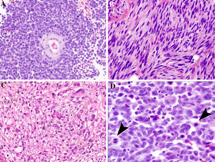
Fig. 2.
Morphologic features in mucosal melanoma. Note that pigmentation is not observed in these examples. a The perivascular/peritheliomatous pattern shows tumor cells loosely clinging around a blood vessel. b Spindled cells create a fasciculated pattern of growth of mimicking other neuronal and soft tissue tumors. c A solid sheet of predominantly clear tumor cells show occasional scattered rhabdoid forms. d Epithelioid cells show characteristic prominent nucleoli and mitoses (arrows)

