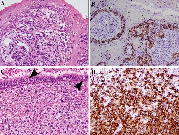
Fig. 3.
Intraepithelial involvement in mucosal melanoma and immunohistochemical confirmation of lineage. a Atypical cells are mixed with acute inflammation in this oral biopsy. b An S100 protein immunohistochemical stain on part A highlights the in situ component and invasive tumor nests. c Pagetoid spread may be identified when the surface mucosa is intact. Large atypical melanocytes (arrows) percolate into the overlying respiratory epithelium. Notice that the underlying tumor is without pigmentation or prominent nucleoli. d An immunohistochemical cocktail of melanoma markers (tyrosinase and HMB45) in case C, strongly highlight the tumor cells confirming tumor lineage

