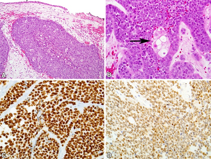
Fig. 4.
NUT carcinoma. a NUT carcinoma grows as nests of tumor cells in the sinonasal submucosa, without a surface epithelial component. a neutrophilic infiltrate is seen. b Most cases of NUT carcinoma demonstrate focal squamous differentiation (arrow) in an abrupt pattern. c NUT carcinoma is usually positive for p40. (D) The diagnosis of NUT carcinoma can be confirmed with diffuse immunoreactivity for NUT protein, typically with a distinctly speckled pattern

