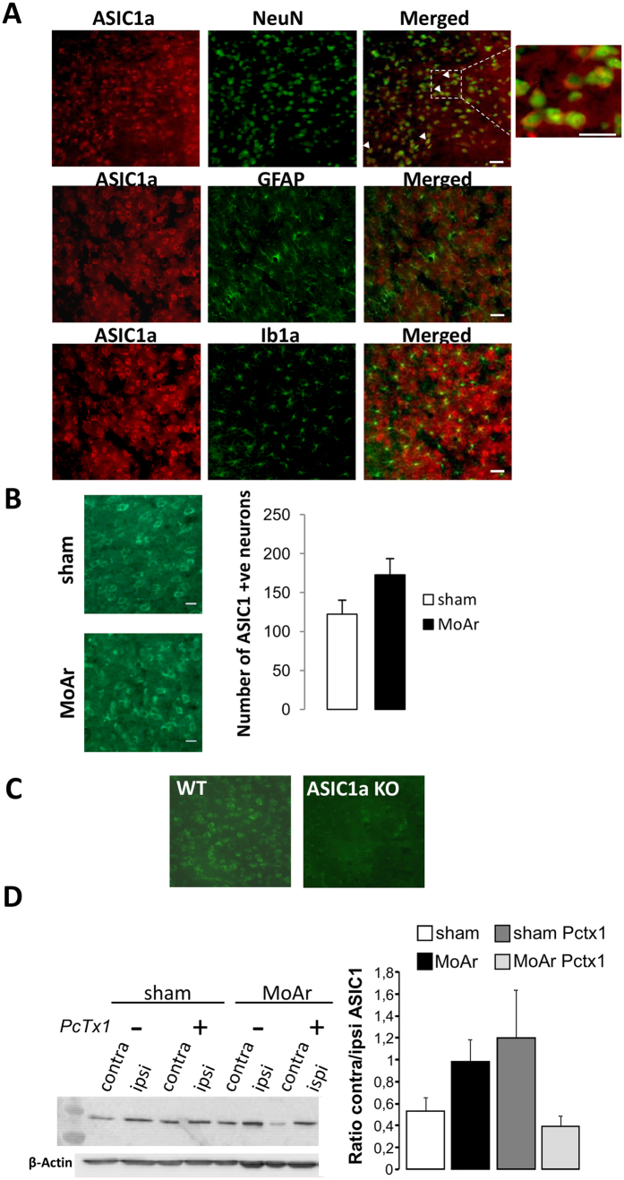
Figure 4. Expression of the ASIC1a protein in the basolateral amygdala (BLA) of MoAr rats.
(A) And inset. ASIC1a (rabbit anti-ASIC1a, 1:500, alpha diagnostic) co-localized with the neuronal marker NeuN (upper panel, mouse anti-neuronal nuclei (anti-NeuN); 1:1,00) but not with an astrocyte marker (GFAP, middle panel, mouse anti-glial fibrillary acidic protein (anti-GFAP); 1:1,000) or a microglial marker (Ib1a, lower panel, rabbit anti-ionized calcium binding adaptor molecule 1 (anti-Iba1); 1:200) in MoAr rats. (B) Quantification of the number of ASIC1a-positive neurons in the contralateral BLA between sham and MoAr animals (n = 5 per group). (C) ASIC1a immunostaining (rabbit anti-ASIC1a, 1:500, alpha diagnostic) in the amygdala of wild type (left panel) and ASIC1a knockout mice (right panel). (D) Representative Western blot of the expression of the ASIC1a protein in ipsilateral and contralateral BLA of sham and MoAr rats treated with saline or PcTx1 (β-actin detection used for normalization shown on the bottom). Densitometric quantification of the signal corresponding to ASIC1a relative to β-actin and expressed as the ratio between the ipsi- and contralateral side for each group. n = 5 per group. One-way ANOVA followed by a Tuckey post hoc test.

