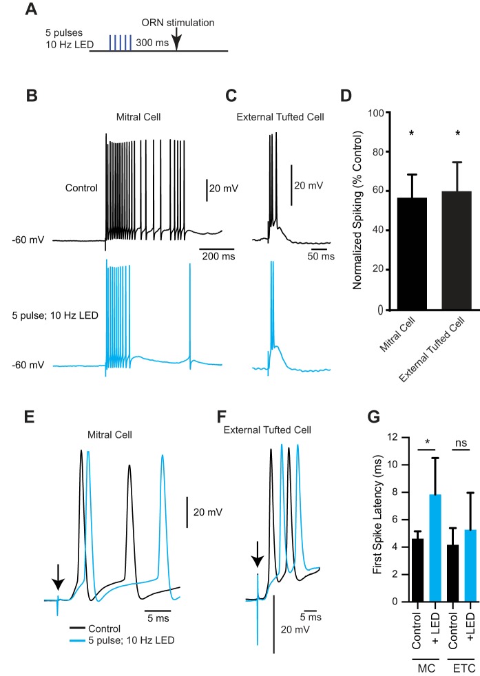
Fig. 4.
Presynaptic inhibition of mitral cell and external tufted cell afferents reduces spiking. A: optogenetic protocol: 5 LED pulses at 10 Hz followed by ORN stimulation (300-ms ISI). B: mitral cell spiking induced by ORN stimulation in control condition (top, black) and after short axon cell activation (bottom, blue). C: external tufted cell spiking induced by ORN stimulation in control condition (top, black) and after short axon cell activation (bottom, blue). D: optogenetic stimulation of short axon cells significantly reduced the ORN-evoked spiking in both mitral and external tufted cells. E and F: higher temporal resolution of the first couple of spikes elicited in control (black) and after LED stimulation (blue) in mitral cells (E) and external tufted cells (F). G: optogenetic stimulation of short axon cells significantly increased the first spike latency in mitral cells (MC) but not external tufted cells (ETC); *P < 0.05..

