
Fig. 7.
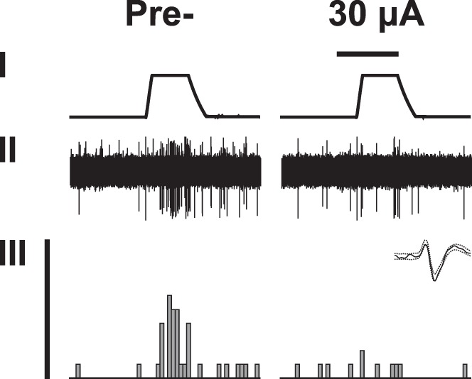
An example of original recording of a wide dynamic range neuron in the spinal dorsal horn of an animal with neuropathic hypersensitivity. Trace I shows the temperature of the contact thermode applied to the hind paw of the nerve-injured limb (baseline 35°C, peak 53°C). Trace II shows raw data of neuronal recording. Trace III shows the peristimulus time histogram, above which is shown the template of the recorded WDR neuron. In the left column, recording before S2 stimulation (Pre-). In the right column, recording during stimulation of S2 contralateral to the tested limb. Horizontal bar above the temperature trace represents 15 s and indicates the duration of S2 stimulation at the intensity of 30 µA.