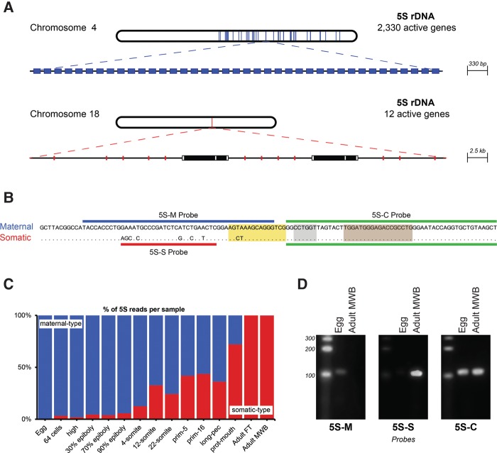
FIGURE 1.
5S rRNA variant types and their expression in zebrafish. (A) Chromosomal localization and organization of the maternal-type (blue) and somatic-type 5S (red) rRNA main clusters in the zebrafish genome. The colored parts on the chromosomes indicate the areas with 5S rRNA genes. In the zoomed-in parts, each colored rectangle is an individual 5S rRNA gene and each white rectangle is a MutsuDrS1 retrotransposon with two potential ORFs (black). (B) Sequence comparison of the 5S rRNA variant types. Maternal, Seq1 sequence; somatic, Seq87 sequence. Internal control regions are shown as colored boxes. A box (yellow); internal element (gray); C Box (light brown) (Schramm and Hernandez 2002; Martins and Wasko 2004). The probes used for Northern blotting are indicated with colored lines. (C) Expression of the 5S rRNA maternal and somatic types indicated by percentage of total 5S rRNA sequencing reads. (Adult FT) Adult female tail; (Adult MWB) adult male whole-body. (D) Northern blot analyses with total RNA from zebrafish eggs and adult (male) tissue and probes as indicated in B. Probe 5S-M recognizes a sequence specific for the maternal-type 5S RNA; probe 5S-S a sequence specific for somatic-type 5S RNA; and probe 5S-C a common sequence between the two 5S RNA variants. Each panel contains lanes from the same gel. In the second panel, the contrast was adjusted as the LNA 5S-S probe gave a very bright signal.

