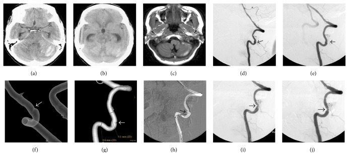
Figure 2.
CT (a) showed lamellar infarct in the pons and worm of cerebellum and SAH. CT (b) showed obstructive hydrocephalus. MRI (c) showed SAH gradually decreased after ventriculoperitoneal shunt. Lateral and oblique DSA ((d) and (e)) of left VA showed the dissection in the segment of V2. Transparency and volume rendering ((f) and (g)) showed the intimal flap. It is the major appearance. During the procedure, the micro-guide wire was difficult to pass through the true lumen until the microcatheter was used (h and i). Lateral DSA showed slight remnant stenosis after stenting (j). CT: computed tomography; MRI: magnetic resonance image; DSA: digital subtraction angiography; VA: vertebral artery.

