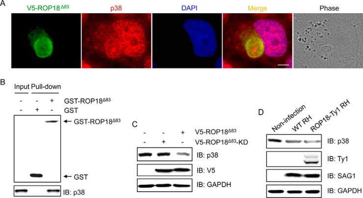
Fig. 5.
ROP18 interacts with p38 and promotes its degradation. A, HeLa cells transiently expressing V5-ROP18Δ83 were infected with tachyzoites of RH strain. 24 h after infection, immunofluorescence staining was performed with mouse monoclonal anti-V5 (green) and rabbit monoclonal anti-p38 (red) antibodies. DAPI (blue) was used to visualize cell nuclei. Phase, bright field; bar, 5 μm. B, GST pulldown assay was performed using GST-ROP18Δ83 protein and 293T cell lysates. IB, immunoblot. C, 293T cells were transfected with V5-ROP18Δ83 or V5-ROP18Δ83-KD plasmids for 36 h, and then total cell lysates were subjected to Western blotting analysis. D, 293T cells were infected without or with wild-type (WT) RH or ROP18-Ty1 RH strain for 36 h. Cellular lysates were separated by SDS-PAGE and immunoblotted with the indicated antibodies. SAG1 and GAPDH were used as loading controls.

