Fig. 3.
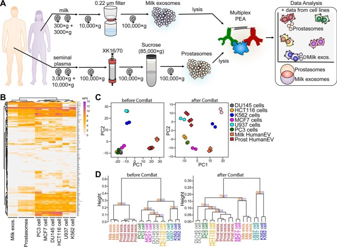
Comparison of protein profiles in lysates from cell lines and in body fluid-derived exosomes reveals tissue origin of exosomes in milk and seminal fluid. A, Illustration of experimental layout. Briefly, breast milk-derived exosomes and prostasomes isolated from healthy donors were submitted to multiplex PEA, and further compared with six cell line lysates (see Fig. 2). B, Heatmap for proteins detected in U937, K562, HCT116, DU145, MCF7 and PC3 cell line lysates together with milk exosomes and prostasomes. C, PCA comparing samples before and after removal of cell- or exosome-specific proteins using ComBat analyses. D, Hierarchical clustering for cell lysates and body fluid-derived exosomes before and after removal of cell- or exosome-specific proteins with ComBat. Protein levels are shown as NPX values. For dendrogram, heights represent dissimilarity among clusters and numbers on the plot indicate approximate unbiased p value (orange) and bootstrap probability (purple). Two to five biological replicates were analyzed for each sample type.

