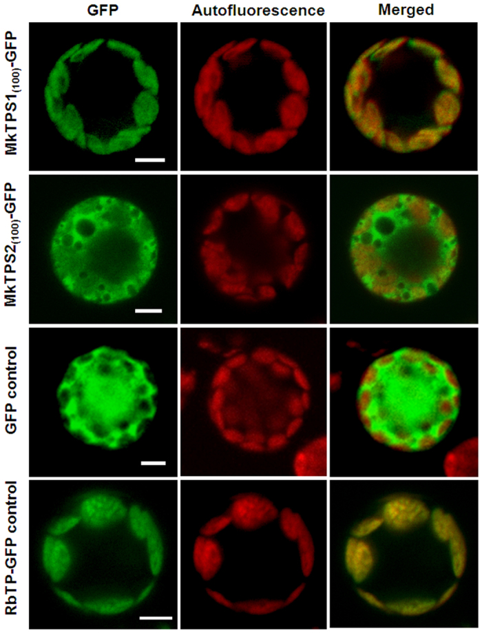
Figure 5. Subcellular localization of M. koenigii terpene synthases.

Confocal laser scanning microscopy of MkTPS1 and MkTPS2 using GFP-fusion proteins in Arabidopsis protoplasts. Name of GFP fusion constructs are shown on the left, and the corresponding transient expression in the protoplasts is shown on the right. GFP fluorescence detected is shown in the ‘GFP’ column; chlorophyll autofluorescence is shown in the “Autofluorescence” column, and the “Merged” column shows combined GFP fluorescence and chlorophyll autofluorescence. Scale bars = 50 μm. The numbers in the fusion constructs correspond to amino acid positions. RbTP (plastidial RuBisCO target peptide)-GFP is a chloroplast marker.
