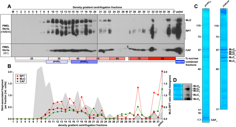
Figure 1. Isolation of PMEL fibrils.
(A) PMEL fibril enrichment via velocity gradient centrifugation coupled with subsequent Triton X-100 extraction of all fractions. Triton X-100-insoluble material from each fraction was analyzed by Western blot. (B) Band intensities in (A) were determined densitometrically (lines). MαC:RPT ratios are shown as filled histogram. (C) SDS-PAGE and Coomassie Blue staining of protoΙΙ and mature fractions. (D) A mature fraction analyzed by Western blot and Coomassie Blue staining.

