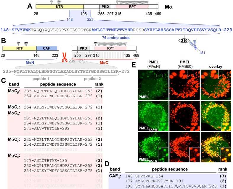
Figure 2. Identification of the PMEL core amyloid fragment.
(A) The mass spectrometry-identified CAF mapped onto the Mα domain structure. The CAF largely overlaps with an N-terminal region, previously referred to as NTR13, but not with the PKD domain. Identified CAF-derived peptides are shown in blue. (B) MαN and MαC domain structure as determined by mass spectrometry. Identified MαC-derived peptides are shown in grey. (C,D) PMEL peptides identified by mass spectrometry in the indicated bands. High confidence peptides (score greater than identity score) shown in black. Lower confidence peptides (score lower than identity score) shown in grey. Peptides are ranked according to score (bold brackets). (E) IF analysis of FlAsH-labeled Mel220 cells stably expressing wt or tetracysteine-tagged PMEL. Antibody HMB50 labels mature fibrils.

