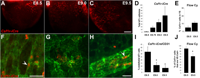
Figure 1. EMPs and EMP-derived cells increase with developmental stage but lose their endothelial cell characteristics at later stages.
Csf1riCreRosa26tdT yolk sacs were harvested at E8.5 (10 somite pairs, A) E9.0 (15 somite pairs, B) and E9.5 (25 somite pairs, C) to visualize tdT+ cells. The number of total of tdT+ cells increased based on immunofluorescence of Csf1riCreRosa26tdT embryos (D, n = 4–5 per stage) and of CSF1R+ cells based on flow cytometry of wild type embryos (E, n = 6 at E8.5 and n = 9 at E9.5). Blood vessels were counterstained with CD31 (F–H, green). Endothelial-like tdT+ cells (F and H, arrowheads) were blindly quantified based on 3 phenotypic criteria (CD31 co-expression, cell shape, and vessel integration) by immunofluoresence. Analysis was limited to yolk sac vessels proximal to the embryo proper. The number of tdT+ cells with an endothelial phenotype was initially 32% but decreased in older embryos (I, n = 4–5 embryos per stage). The percentage of CSF1R+ cells that co-stained for VE-Cadherin was also measured by flow cytometry and confirmed the decrease in endothelial staining in older embryos (J). Scale bars: 250 μm (A–C), 50 μm. (F–H) All values are mean ± SEM. *p < 0.05; two-tailed Student’s t-test.

