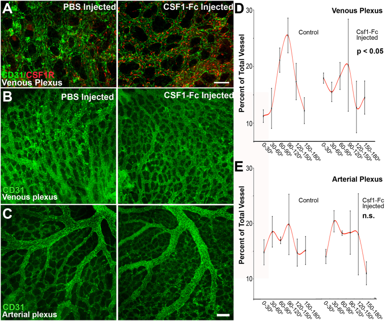
Figure 7. Exogenous CSF1-Fc administration increases the number of early myeloid cells and disrupts remodeling in the venous plexus.
Embryos were dissected at E8.5 (10 somite pairs) and injected with either PBS or CSF1-Fc and then cultured for 24 hours. CSF1-Fc treatment resulted in an increase in the number of CSF1R+ cells (red) localizing to the venous plexus (A, n = 5–6 embryos). Injection of CSF1-Fc disrupted venous (B), but not arterial remodeling (C). Blinded quantification of vessel angles confirms that remodeling was significantly impaired in the venous plexus (D), but not arterial plexus (E). Scale bars: 100 μm (A), 200 μm (B,C). All values are mean ± SEM. Distributions were compared using a Kolmogorov-Smirnov test.

