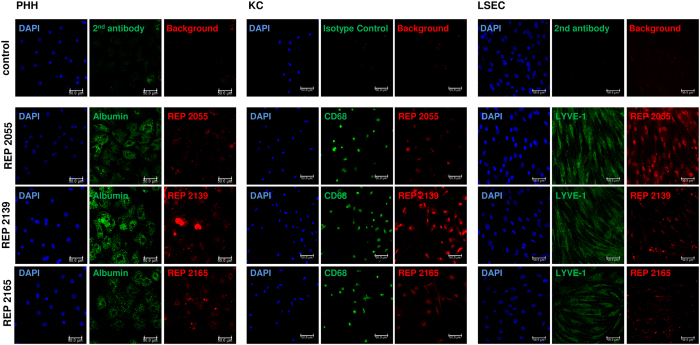
Figure 2. Nucleic acid polymers (NAPs) were efficiently taken up by different liver cell types.
The identity of primary human hepatocytes (PHH) (A), Kupffer cells (KC) (B) and liver sinusoidal endothelial cells (LSEC) (C) was assessed by immunofluorescent staining of cell type-specific markers albumin (A), CD68 (B) and LYVE-1 (C) (green), respectively. Nuclei were counterstained with DAPI (blue). Uptake of NAPs was visualized using Cy3-labeld (red) REP 2055 [0.01 μM], REP 2139 [0.05 μM] and REP 2165 [0.05 μM]. Immunofluorescence staining was detected with a laser scanning microscope (LSM; Axiovert 100 M; Zeiss, Jena, Germany) at 20 × magnification. Image analysis was performed with LSM Image Browser (Zeiss). Scale bar 50 μm.

