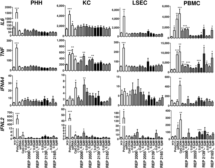
Figure 3. Cell type-specific expression of innate immune genes in response to NAP treatment in vitro.
Primary human hepatocytes (PHH, n = 3–5), Kupffer cells (KC, n = 3–5), liver sinusoidal endothelial cells (LSEC, n = 3–6) and peripheral blood mononuclear cells (PBMC, n = 3–6) were stimulated with DNA-based (REP 2006 and REP 2055) and RNA-based (REP 2139 and REP 2165) NAPs or immunostimulatory controls (TLR3 agonist Poly(I:C); TLR7/8 agonist ssRNA40 [ssRNA] and TLR9 agonist CpG ODN2216 [CpG ODN]) for 6 h. RNA was extracted, and gene expression of interleukin 6 (IL6), tumor necrosis factor (TNF), interferon alpha 4 (IFNA4) and interferon lambda 2 (IFNL2) was assessed by quantitative reverse transcription polymerase chain reaction (qRT-PCR). Values represented mean ± SEM (normalized to 100,000 copies of beta actin (ACTB) mRNA). Group size n = 3–6 cell preparations. Statistically significant changes compared to untreated controls are reported for p < 0.05 (*), p < 0.01 (**), p < 0.001 (***); w/o, without treatment.

