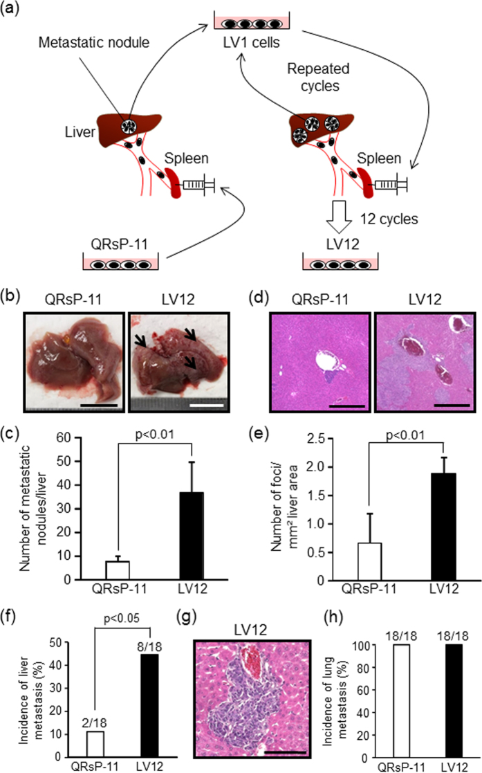
Figure 1. LV12 cells possess a high liver-metastatic potential and give rise to multiple liver colonies by intrasplenic and intravenous injections.
(a) Schematic representation of in vivo sequential selection of a liver-metastatic variant subline (LV12) from QRsP-11 mouse fibrosarcoma cells. Metastatic nodules in the livers of C57BL/6 mice, which formed from 1 × 10⁶ QRsP-11 cells previously injected into the spleen, were harvested and a cell culture subline was established. These cells were then injected into the spleens of other mice, and liver metastases subsequently formed. The metastatic foci were excised and expanded as a new subline. This procedure was repeated 12 times, yielding a cell subline designated as LV12. (b) Macroscopic views of liver metastasis formation 7 days after intrasplenic injection of 1 × 10⁶ LV12 or QRsP-11 cells. Arrows indicate representative metastatic nodules. Scale bar: 10 mm. (c) The numbers of metastases on the liver surface were determined. Bar graphs show means ± SD (n = 5 in each group). (d) Representative H&E staining of the livers from Fig. 1b. Scale bar: 500 μm. (e)The number of metastatic foci per mm2 of liver area was determined using image analysis software. Bar graphs show means ± SD (n = 5 in each group). (f) The incidence of liver metastatic colonization 7 days after tail vein injection of 1 × 10⁶ LV12 or QRsP-11 cells was evaluated using histology. Bar graphs show means ± SD from three independent experiments with similar results (n = 18 in each group). (g) Microscopic appearance of the liver metastatic foci formed by LV12 cells. Scale bar: 100 μm. (h) The incidence of lung metastasis was determined by the presence of metastatic nodules on the lung surface on day 7 post-injection. The incidence of metastatic colonization is indicated above each bar (number of mice with metastasis/number of mice examined).

