
Figure 7. Location of the putative cholesterol binding sites on the predictive 3D model of LAT1.
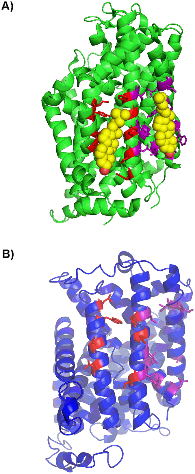
(A) Crystal structure of dDAT (PDB: 4xpf) bound to cholesterol (yellow and red spheres) (B) Predictive 3D structure of LAT1 generated by the I-TASSER server and optimised to improve backbone stereochemistry. Key residues in the cholesterol/CHS binding sites I (purple) and II (red) of dDAT and the corresponding residues in the putative sites of LAT1 are shown as sticks.