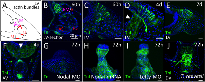
Figure 3. Expression of HpTnI protein in sea urchin larvae.
(A) Schematic of muscular actin bundles in sea urchin larva. M, E, EM, PS, and AS indicate mouth, esophagus, esophageal muscles, pyloric sphincter and anal sphincter, respectively. (B) An optical section of a 60-h larva expressing HpTnI in the esophagus, esophageal region and the regions of the pyloric (PS) and anal (AS) sphincters. (C) Stacked image of a 60-h larva. HpTnI patterns are similar between 4 days (D) and 7 days (E). HpTnI expression in the ventral ectoderm is conspicuous by 4 days (D, arrowhead). (F) The anterior of the larva is at the top. HpTnI expression in the ventral ectoderm is clearly visible (arrowhead). (G) No ectodermal expression of HpTnI is visible in a Nodal morphant. (H and I) By contrast, with Nodal overexpression and in a Lefty morphant, the whole ectoderm except for the anterior and posterior ends expresses HpTnI. (J) A similar TnI pattern was observed in 72-h larvae of Temnopleurus reevesii. LV, lateral view; AV, anal view; DV, dorsal view. Bar = 20 μm.

