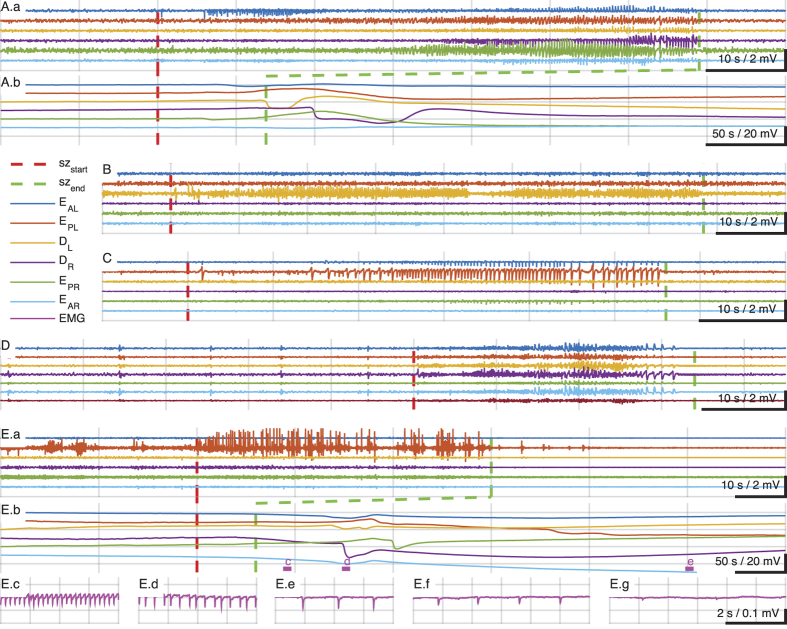
Figure 4. Examples of seizure types and SUDEP.
Seizures were classified using the 2010 International League against Epilepsy seizure classification system18 depending on the mode of onset and semiology using video-EEG recordings. (A.a) Seizure of cortical onset with secondary generalization. In compressed time-scale below, (A.b), note direct current (DC) potential changes in cortical and hippocampal electrodes consistent with propagating spreading depression (SD) following seizure termination (vertical broken green line). In (B) is shown a focal hippocampal seizure, and in C a focal cortical seizure, both from the same animal. (D) Illustrates an example of a primary generalized seizure preceded by a series of pre-ictal generalized spikes. In (E) is shown an example of a sudden death during seizure. The cortical focal seizure shown in (E.a) is punctuated by the animal becoming behaviorally quiet, and is followed by propagating depolarizations consistent with SD, shown in (E.b). Following the seizure, the muscle activity is quiet enough to reveal the EKG reflected in the EMG lead which demonstrates progressive bradycardia leading to asystole shown in (E.c–E.g). EEG montage: EAL, EEG anterior left (frontal); EPL, EEG posterior left (somatosensory); DL, depth hippocampus left; DR, depth hippocampus right; EAR, EEG anterior right, EPR, EEG posterior right. Filter settings for traces shown: Seizure traces bandpass 1–50 Hz; SD traces low-pass below 1 Hz; EMG/EKG traces 0.1–55 Hz.

