Fig. 3.

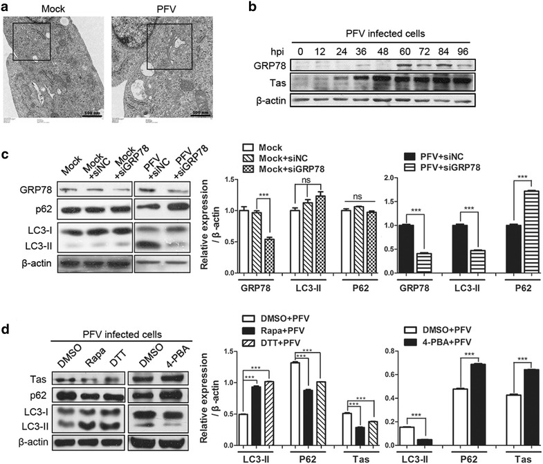
PFV triggers autophagy by activating ER stress. a Expansion of the ER was detected via transmission electron microscopy (TEM). BHK-21 cells infected with mock supernatant or PFV at an MOI of 0.5 were processed and analyzed at 24 hpi for the ER expansion via electron microscopy. Black frames indicated representative expanded ER lumen. Scale bars 500 nm. b BHK-21 cells were infected with PFV to analyze GRP78/Bip protein expression by western blotting. c BHK-21 cells were transfected with the siRNAs of GRP78 or siRNAs of Negative control for 36 h, followed by infection with mock supernatant or PFV at an MOI of 0.5 or mock infecting. After 1.5 h of virus absorption at 37 °C, the cells were further cultured in maintenance medium. At 24 h after infection with mock or PFV, the cells were subjected to western blotting using anti-GRP78, anti-P62 and anti-LC3 and antibodies. In lane 1, cells were only infected with mock supernatant and cultured in maintenance medium for 48 h. d Western blotting was used to analyze the expression of the viral protein Tas and LC3 in PFV-infected cells in the absence or presence of the ER stress inducer DTT (1 mM) or the ER stress inhibitor 4-PBA (1 mM). Rapa-treated (400 nM) cells were used as a positive control. Significance was analyzed with a two-tailed Student’s t test. ns P > 0.05, ***P < 0.001
