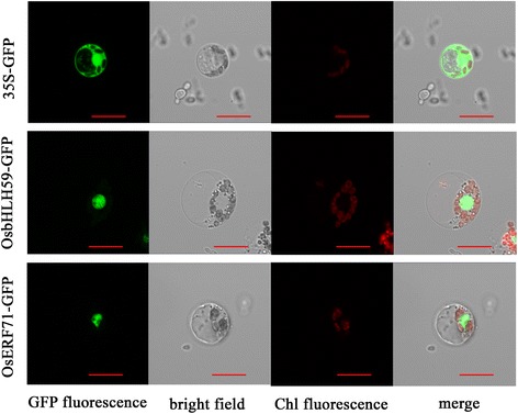
Fig. 6.

Subcellular localization of OsbHLH59 and OsERF71 in rice protoplasts. The rice protoplasts were transformed with 35Sp::OsbHLH59:GFP, 35Sp::OsERF71:GFP or pCAMBIA1300-GFP. The transformed cells were observed under a confocal microscope. The photographs were taken under detecting GFP fluorescence, bright field, chloroplast auto-fluorescence, and merged microscope images, respectively. Empty vector (pCAMBIA1300-GFP) was used as a control. Bars, 10 μm
