
Figure 3.
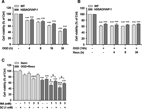
Effect of OGD, OGD with re‐oxygenation and MA on cell viability of WT and human SSAO/VAP‐1‐transfected hCMEC/D3 cells [hCMEC/D3 hSSAO/VAP‐1]. Different durations of OGD (A) or 16 h OGD with different durations of re‐oxygenation (B) induced the death of WT and hSSAO/VAP‐1‐transfected hCMEC/D3 cells. MTT reduction assay was used to assess cell viability under the different assay conditions. Cells without OGD were maintained under normoxic conditions and were considered to be control cells (Ctrl). The metabolism of MA subjected to 16 h OGD plus 24 h of re‐oxygenation (OGD + Reox) (C) induced a reduction in cell viability in hSSAO/VAP‐1‐expressing hCMEC/D3 cells, which was partially prevented by the SSAO activity inhibitor SC. MA (1 or 3 mM) and SC (1 mM) were added before OGD and maintained during re‐oxygenation. Control cells are untreated cells in normoxia (Norm). Data are expressed as mean ± SEM of at least three independent experiments. *** P < 0.001 versus control of the corresponding cell type. # P < 0.05 and ### P < 0.001 versus untreated hSSAO/VAP‐1‐expressing hCMEC/D3 cells in the corresponding condition (Norm or OGD + Reox); & P < 0.05 between the indicated treatments. Statistical analyses were performed by a one‐way ANOVA and the addition of Newman–Keuls multiple comparison test.