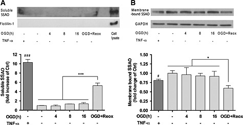
Figure 4.

OGD with re‐oxygenation induces the release of soluble SSAO to the culture media by hCMEC/D3 hSSAO/VAP‐1 cells. (A) Levels of soluble hSSAO/VAP‐1 in 10‐fold concentrated culture media, in response to TNF‐α treatment (24 h in normoxia, 100 ng · mL−1), different OGD times and 16 h OGD with 24 h re‐oxygenation (OGD + Reox). TNF‐α was used as a positive control of SSAO/VAP‐1 release. Flotillin‐1 was used as control of cell debris absence in the media. (B) Presence of membrane‐bound SSAO/VAP‐1 in hCMEC/D3 hSSAO/VAP‐1 cell lysates under the same experimental conditions as in (A). The presence of membrane‐bound SSAO was normalized to the GAPDH levels. Untreated cells or media under normoxia conditions were considered control samples (Ctrl). Data in graphs represent the quantification of Western blots and are expressed as mean ± SEM of data obtained from three independent experiments. * P < 0.05 and *** P < 0.001 as indicated; # P < 0.05 and ### P < 0.001 versus control cells, by a one‐way ANOVA and the addition of Newman–Keuls multiple comparison test.
