Figure 1.

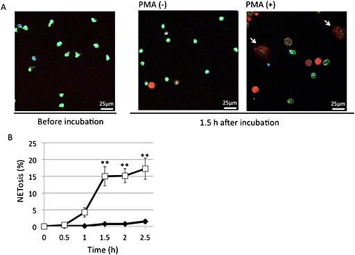
PMA‐induced NETosis in isolated neutrophils in vitro. After human peripheral neutrophils were incubated at 37°C for 1.5 h, without or with 50 nM PMA, they were stained with cell‐permeable DNA stain Hoechst 33342 (blue), mitochondrial stain mitotracker green FM (green) and cell‐impermeable DNA stain sytox orange (red) and analysed by confocal microscopy. (A) Typical photos with arrows indicating neutrophils undergoing NETosis. (B) Time‐dependent increase of cells undergoing NETosis. NETosis was visually measured in more than 300 cells in five randomly selected fields by confocal microscopy. Data are expressed as mean percentage of neutrophils undergoing NETosis per total number of cells ± SEM of five independent experiments. **P < 0.01 compared with the PMA (−) sample at each time point.
