
Figure 6.
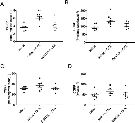
BoNT/A effect on concentration of CGRP protein in dura, trigeminal nucleus caudalis, trigeminal ganglion and CSF in CFA‐treated rats. The 5 U kg−1 BoNT/A was injected into the TMJ 3 days before the CFA treatment. Tissues were collected 1 day post‐CFA, and the CGRP concentration was analysed by RIA. (A) Dura mater; (B) ipsilateral trigeminal nucleus caudalis; (C) ipsilateral trigeminal ganglion; and (D) CSF. Scatter plot represents individual animal values, and horizontal lines and bars indicate mean ± SEM. n (animals per group) = 6. *P < 0.05, significantly different from saline control; **P < 0.01, significantly different from saline control; ++ P < 0.01, significantly different from saline + CFA; one‐way ANOVA followed by Newman–Keuls post hoc test.