
Figure 7.
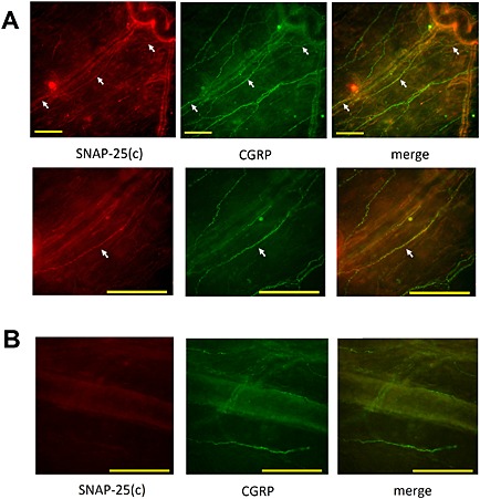
Colocalization of truncated SNAP‐25 and CGRP in ipsilateral cranial dura after BoNT/A injection in the periphery. BoNT/A 20 U kg−1 total dose was injected into four different sites (TMJ, whisker pad, and frontal and temporal regions; 1.75 U/20 μL per site) on the left side of the head. Animals were perfused for immunohistochemistry 6 days later. (A) Upper panel: lower magnification fluorescent microphotograph shows the course of a single‐cleaved SNAP‐25 [SNAP‐25(c)]‐immunoreactive fibre (arrows, red immunofluorescence) in the vicinity of dural blood vessels, which colocalizes with CGRP (green fibers). Lower panel: higher magnification image of the middle part of cleaved SNAP‐25‐immunoreactive fibre, which colocalizes with granular CGRP immunofluorescence. (B) Microphotograph of contralateral side dura of the same animal without detectable cleaved SNAP‐25 in CGRP‐expressing afferents. The images are representative of the data obtained from four animals. Scale bars = 100 μm.