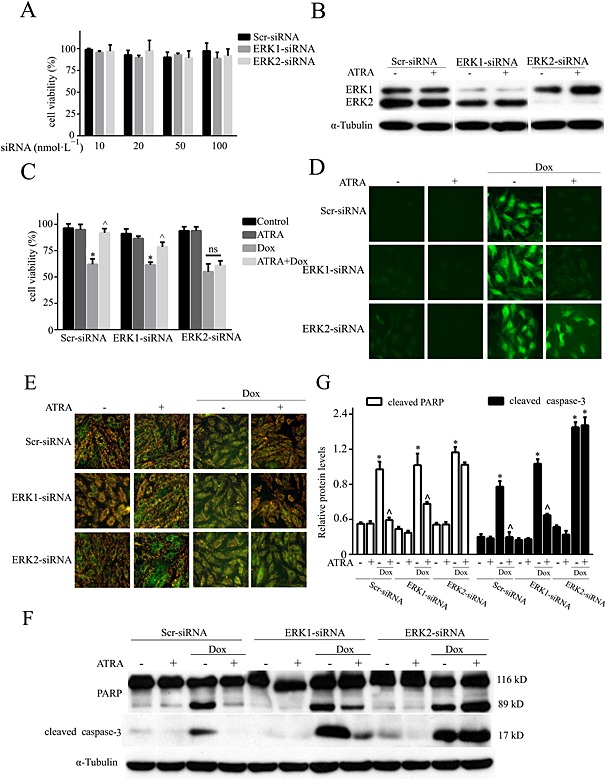
Figure 7.

The role of ERK1 and ERK2 knockdown in the ATRA‐mediated protective effect on doxorubicin cardiotoxicity. (A) Toxicity of ERK1 and ERK2 siRNA in H9c2 cells; (B) silencing of ERK1 and ERK2 and ERK expression and activation under basal and ATRA‐stimulated conditions. Effects of ERK1 and ERK2 silencing on (C) cell viability, (D) ROS (DCFH‐DA staining), (E) MMP (JC‐1 staining) and (F) the levels of apoptosis‐associated proteins (Western blotting) in ATRA cardioprotection (n = 5). (G) Semi‐quantitative results for apoptosis‐associated proteins (cleaved PARP and cleaved caspase‐3). Quantitative values are computed as the ratios of the density of the targeted protein to that of α‐tubulin. Except as noted, the concentrations of ATRA and doxorubicin were 2 and 3 μmol·L−1 respectively. Data are expressed as the mean ± SEM and were analysed by anova. *P < 0.05 versus control and ^P < 0.05 versus Dox.
