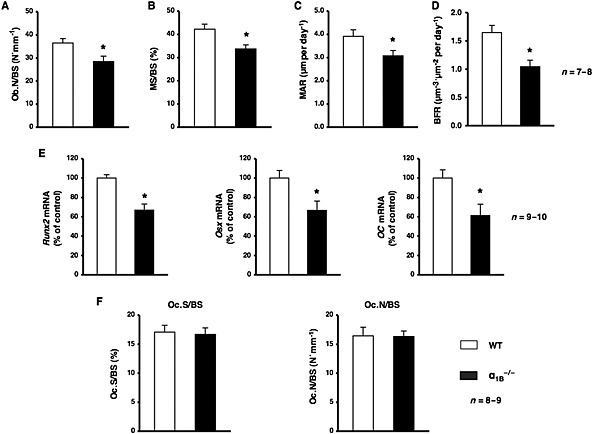
Figure 4.

Ablation of α1B‐adrenoceptors led to a decrease in bone formation. (A–D) An analysis of the Ob.N/BS, MS/BS, MAR and BFR in the cancellous bone compartment of the distal femur metaphysis from WT mice and α1B −/− mice; n = 7 or 8 mice per group. (A) Ob.N/BS. (B) MS/BS. (C) MAR. (D) BFR. Values are expressed as means ± SEM. * P < 0.05, significantly different from WT mice. (E) Total RNA was isolated from the distal region of the femur from WT and α1B −/− mice, followed by the determination of Runx2, Osx and OC mRNA levels by real‐time qRT‐PCR using specific primers; n = 9 or 10 mice per group. Values are expressed as means ± SEM. * P < 0.05, significantly different from WT mice. (F) Sections of the primary trabecular regions of the femur from WT or α1B‐adrenoceptor‐deficient mice followed by the staining of osteoclasts with TRAP. The amount of Oc.S/BS and Oc.N/BS were measured (n = 8 or 9 mice per group).
