
Figure 5. Ablation of BMP9 increases the number of vessel junctions and reduces lacunarity in primary tumor vasculature.
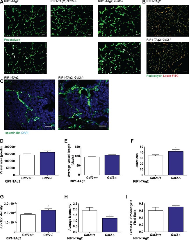
(A) Representative images of Podocalyxin-stained vessels in PanNETs of 12-week old RIP-TAg2 mice. Vessel junctions are highlighted in second row with white dots. Scale bar 20 μm. (B) Representative images of Lectin-FITC perfused vessels in PanNETs of 12-week old RIP-TAg2 mice. Scale bar 50 μm. (C) High-magnification vessel details in PanNETs stained with Isolectin GS-IB4. Scale bar 20 μm. (D–H) Analysis of vessel features in Podocalyxin-stained PanNETs from RIP1-TAg2 mice; total vessel area (C), average vessel length (D), total number of junctions (E), junction density (F), and lacunarity (G). (I) Quantification of immunofluorescence of FITC-conjugated Lectin perfusion relative to Podocalyxin expression. Data are mean ± SEM. *P < 0.05 vs. Wildtype with Student's t-test.