
Figure 1.
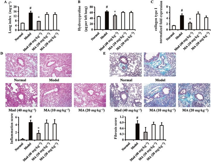
Effects of madecassoside (Mad) and madasiatic acid (MA) on BLM‐induced PF in mice. (A) Body wt (g) of each mouse was recorded, and lung wet wt (mg) was determined immediately after it had been removed. The lung index (mg·g−1) was calculated by dividing the wet lung wt by the body wt. (B) The hydroxyproline concentrations in the upper lobes of left lung tissues were measured by using kits according to manufacturer's instructions. (C) The mRNA expression of collagen type I in the right lung tissues was measured by using Q‐PCR assay. The relative expression of transcription factors was normalized to that of GAPDH in each sample. (D, E) Histopathological changes in the lower lobes of left lung tissues were examined by H&E stain and Masson's trichrome stain (original magnification 200×) respectively. The histological scores of all groups were calculated. Data are expressed as means ± SEM, n = 6–8. # P < 0.05 versus normal; * P < 0.05 versus model.