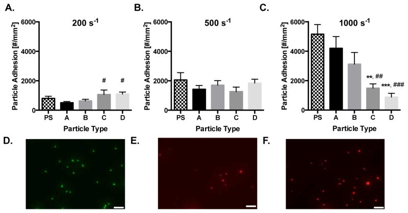
Figure 3. Particle adhesion to inflamed HUVEC monolayer as a function of particle modulus.
Quantified adhesion of 2 μm hydrogel particles at a wall shear rate of (A) 200 s−1, (B) 500 s−1, and (C) 1,000 s−1 by modulus after 5 mins of laminar blood flow over an IL-1β activated HUVEC monolayer. N=3–6 human blood donors per particle condition. Statistical analysis of adherent density was performed using one-way ANOVA with Fisher’s LSD test between all particle adhesion conditions. (*) Represent comparison to PS and (#) represents comparison to particle type A. (*) indicates p<0.05, (**) indicates p<0.01, and (***) indicates p<0.001. Error bars represent standard error. Representative fluorescent images of particles bound to IL-1β activated HUVEC under a WSR of 200 s−1 in vitro for 2 μm (D) PS, (E) Hydrogel A, and (F) Hydrogel D. Scale bars are 20 μm.

