Fig. 1.

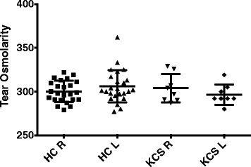
Tear osmolarity studies were done as described in materials and methods. Data shown are the tear osmolarity in the right and left eye of the normal controls (HC) and the patients with keratoconjunctivitis sicca (KCS)

Tear osmolarity studies were done as described in materials and methods. Data shown are the tear osmolarity in the right and left eye of the normal controls (HC) and the patients with keratoconjunctivitis sicca (KCS)