
Figure EV5. Gallbladder organoids prepared from mice expressing constitutively GFP and engraftment in the liver parenchyma.
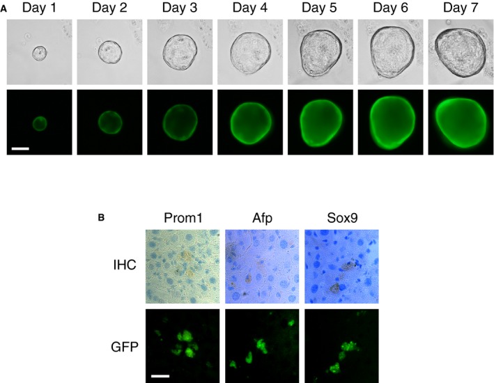
- Phase‐contrast (top panels) and epifluorescence (bottom panels) microscopy images of a representative organoid over a seven‐day period. Scale bar: 200 μm.
- Engraftment of cells isolated from gallbladder organoids in the liver parenchyma. Immunohistochemistry images (top) showing expression of Prom1, α‐fetoprotein (Afp), and Sox9 in cells that express GFP (bottom). Scale bar: 25 μm.