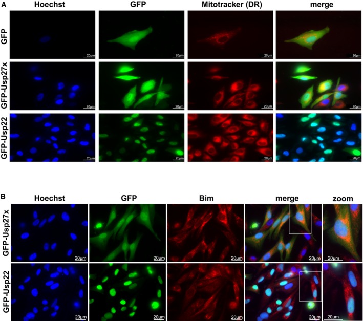
Figure EV4. Subcellular localization of Usp27x and Usp22 as analysed by fluorescence microscopy.

- 1205Lu melanoma cells carrying dox‐inducible GFP, GFP‐Usp27x or GFP‐Usp22 were treated for 24 h with dox. Subcellular localization of GFP or GFP‐fusion proteins (green) was visualized by microscopy. Nuclei were stained with Hoechst (blue), and mitochondria were identified by Mitotracker DeepRed FM staining (red). Scale bar, 20 μm. n = 2. In the merged image for GFP‐Usp22, the Mitotracker signal is not shown. GFP‐Usp27x showed very similar localization in HCC827‐GFP‐Usp27x cells (not shown).
- 1205Lu‐GFP‐Usp27x and 1205Lu‐GFP‐Usp22 were treated for 48 h with dox. Subcellular localization of GFP‐fusion proteins (green) was visualized by microscopy. Nuclei were stained with Hoechst (blue), and Bim was detected by intracellular staining for endogenous Bim (red). A higher magnification of merged figures is shown to the right (zoom). Scale bar, 20 μm. n = 2. Similar results were seen in every visual field analysed.
