
Figure EV3. Analysis of interactions between PALB2 versions and key HDR proteins, as well as immunoblots upon siRNA depletion of BLM and FBH1.
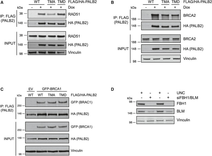
- The PALB2 cell lines were untreated or treated with doxycycline followed by exposure to IR treatment (15 Gy, 2 h recovery). The exogenous FLAG/HA‐tagged PALB2 was immunoprecipitated with FLAG‐agarose and the IP and input samples were run on SDS–PAGE. The membranes were blotted with RAD51 and HA antibodies and vinculin was used as a loading control.
- The cells were treated and analyzed as in (A). Immunoblotting was performed with BRCA2, HA, and vinculin antibodies.
- 293T cells were transfected with empty vector (EV) or GFP‐BRCA1 together with FLAG‐/HA‐tagged WT, TMA, or TMD PALB2. Immunoprecipitation was performed using FLAG‐agarose, and the IP and input samples were probed with GFP and HA antibodies. Vinculin was used as a loading control.
- Representative immunoblot for Fig 3D showing depletion of FBH1 and BLM. The cell lysates were analyzed by Western blotting and probed with FBH1, BLM, and vinculin antibodies.