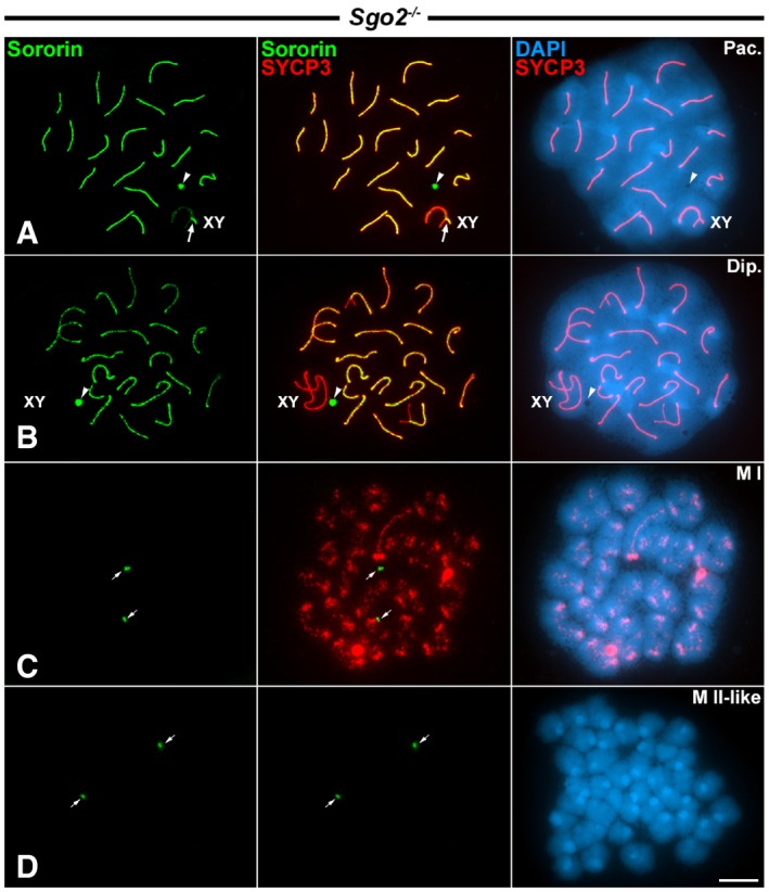
Figure 6. Distribution of Sororin in Sgo2 −/− knockout spermatocytes.

-
A–DDouble‐immunolabeling of Sororin (green) and SYCP3 (red), and counterstaining of the chromatin with DAPI (blue) on spread Sgo2 −/− spermatocytes at (A) pachytene (Pac.), (B) diplotene (Dip.), (C) metaphase I (M I), and (D) metaphase II‐like (M II‐like).
