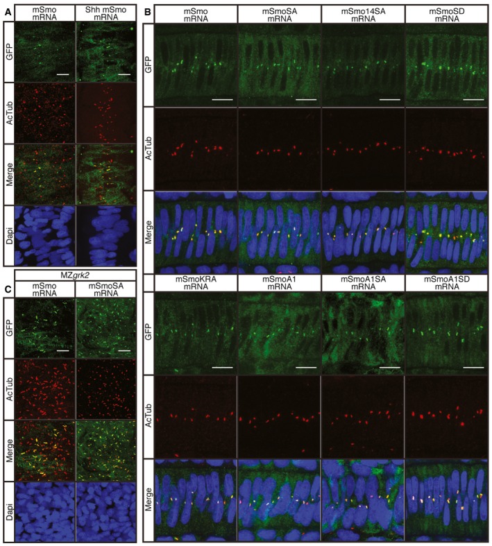
Figure 6. Cilia localisation of wild‐type and mutant Smo.

- Wild‐type 18hpf embryos injected with mRNA encoding GFP‐tagged mSmo (green) showing localisation to the PC of myotomal cells labelled with anti‐acetylated tubulin (AcTub; red), stimulated in response to Shh injection (n = 4). Differences in PC distribution are due to morphological changes in the myotome induced by ectopic expression of Shh as revealed by the distribution of nuclei (DAPI stained; blue). Scale bar, 10 μm.
- Notochord cells of wild‐type embryos expressing GFP‐tagged wild‐type and mutant forms of mSmo. Note the localisation to the PC (labelled with anti‐AcTub; red) in each case (n = 4 for each sample). Scale bar, 10 μm.
- MZgrk2 18hpf embryos injected with mRNA encoding GFP‐tagged wild‐type mSmo or mSmoSA (green) showing localisation to the PC (labelled with anti‐AcTub; red) in myotomal cells. More Smo is localised to the PC in MZgrk2 mutants compared to wild type (panel A). Scale bar, 10 μm.
