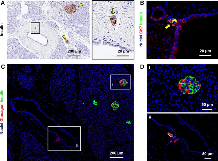
Figure 5.

Expression of pancreatic endocrine markers in pancreatic duct glands (PDGs). (A) Immunohistochemistry for insulin in human pancreas. PDG cells were positive for insulin (arrow in the right image). Area in the box is magnified in the right image. Insulin+ pancreatic islets were present (arrowheads) and represented the positive control. (B) Double immunofluorescence for insulin (green) and cytokeratin7 (CK7: red); the nuclei are displayed in blue. Insulin+ cells within PDGs are CK7+ (arrow). (C,D) Double immunofluorescence for insulin (green) and glucagon (red); the nuclei are displayed in blue. Glucagon+ cells were present within PDGs (magnified in D: arrow). Insulin+ and glucagon+ pancreatic islet cells were present (magnified in D) and represented positive controls. Acinar cells and intercalated ducts were essentially negative for insulin and glucagon.
