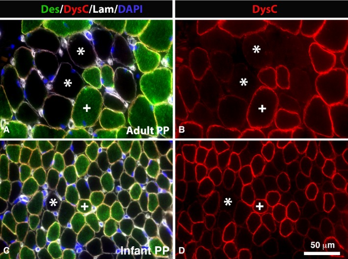
Figure 2.

Des‐Neg and DysC‐Neg fibers in PP. Muscle cross‐sections from an adult (A,B) and infant (C,D) PP muscle multi‐stained for desmin (Des, green color), dystrophin C‐terminal (DysC, red color), laminin (Lam, white color) and DAPI (nuclei, blue color) (A,C) and just for dystrophin C‐terminal (DysC, red color; B,D). Des‐Neg/DysC‐Neg fibers are marked (*) and Des‐Pos/DysC‐Pos are marked (+). Note the high frequency of fibers unstained or weakly stained for both the dystrophin C‐terminal and desmin in adult and infant muscles. Scale bar: 50 μm.
