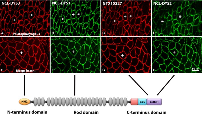
Figure 3.

Immunoreaction of antibodies against different domains of the dystrophin molecule in palate and limb muscles. Serial cross‐sections from palatopharyngeus (A–D) and biceps brachii (E–H) stained with antibodies directed against the N‐terminal (NCL‐DYS3, red color; A,E), the rod (NCL‐DYS1, green color; B,F) and the C‐terminal (GTX15227, red color; C,G and NCL‐DYS2, green color; D,H) domains of the dystrophin molecule. The schematic sketch shows domains of the dystrophin molecule corresponding to antibody affinity. Note that while all domain‐specific antibodies showed immunoreaction in the limb muscle, a subgroup of fibers was unstained or weakly stained for the two antibodies directed against the C‐terminus in the UV (asterisks). Muscle fibers stained for all four antibodies are marked (+). Scale bar: 50 μm.
