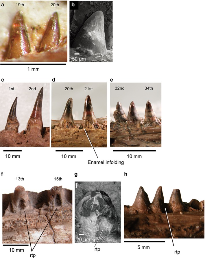
Figure 2.

Marginal and palatal tooth morphology: (a) juvenile Monjurosuchus sp. (IVPP V14261) dentary teeth in medial view; (b) Cteniogenys sp. dentary (UCL uncataloged) in lateral view, SEM; (c) Champsosaurus gigas (SMM P77.33.24) left dentary teeth in lateral view; (d, e) Champsosaurus gigas (SMM P77.33.24) right maxillary teeth in lateral view, image reflected for ease of comparison; (f) Simoedosaurus lemoinei (MNHN BR 1935) replacement maxillary teeth in medial view; (g) Cteniogenys sp. (BMNH R11759), right pterygoid tooth; (h) Champsosaurus sp. isolated right palatine teeth (RTMP 92.36.270) in medial view. Tooth position is uncertain in (b), (g), (h) due to incompleteness of specimens.
