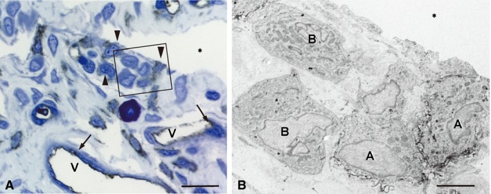
Figure 3.

RECA‐1‐immunoreactivity in the synovial membrane. (A) RECA‐1‐reactions are localized in the endothelial cells (arrows) of the capillaries (V) and several lining cells (arrowheads). (B) An immunoelectron micrograph of the boxed area in (A). Immunopositive lining cells (A) possess filopodia‐like surface folds, indicating macrophage‐like type A cells. Type B cells (B) are immunonegative. *Articular cavity. Scale bars: 20 μm (A), 4 μm (B).
