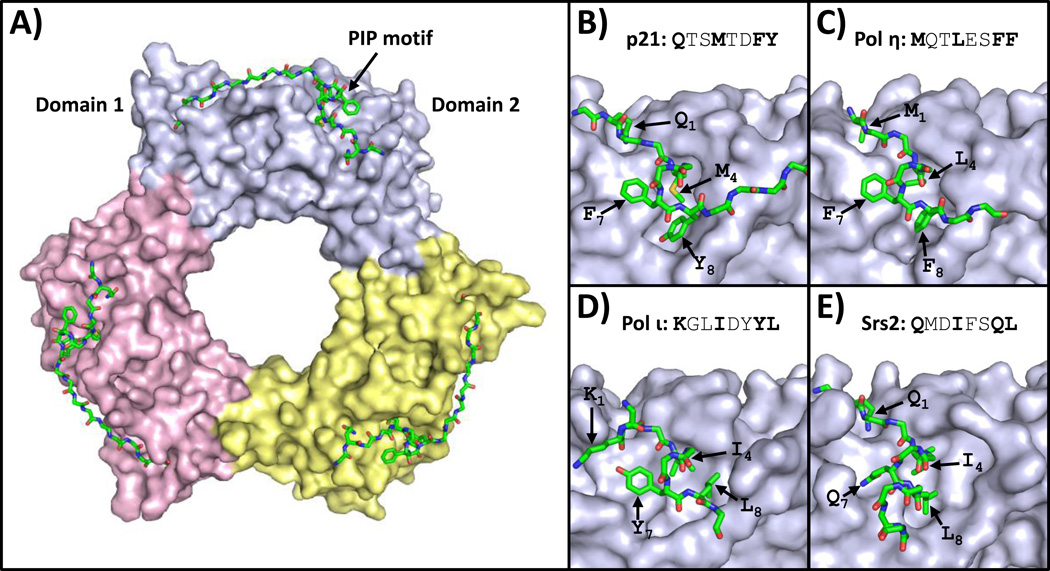
Figure 1.
Structures of PCNA bound to PIP motifs. (A) An overview of the structure of human PCNA bound to peptides containing the PIP motif of p21 (PDB ID: 1AXC) is shown [28]. The three PCNA subunits are depicted in the surface representation (light blue, yellow, and pink). The PIP motif is depicted in the stick representation (with carbon, nitrogen, oxygen, and sulfur atoms colored green, blue, red, and yellow, respectively). (B) A close up view of human PCNA bound to the p21 PIP motif is shown [28]. The side chains of residues in positions 1, 4, 7, and 8 are indicated. (C) A close up view of human PCNA bound to the pol η PIP motif (PDB ID: 2ZVK) is shown [29]. (D) A close up view of human PCNA bound to the pol ι PIP motif (PDB ID: 2ZVM) is shown [29]. (E) A close up view of yeast PCNA bound to the Srs2 PIP motif (PDB ID: SV62) is shown [30].

