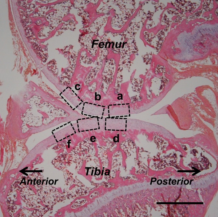
Figure 1.

Regions of assessment used in the histological imaging of sagittal sections (stained with HE after 8 weeks of immobilization) of the knee at the femur (a–c) and tibia (d–f), representing the contact (a, d), transitional (b, e) and non‐contact regions (c, f). Scale bar: 1 mm.
