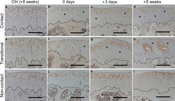
Figure 8.

Immunohistochemical staining to detect CD44 expression in the tibia at 0 days (b, f and j), 3 days (c, g and k) and 8 weeks (d, h and l) after remobilization. Positive cells were observed in most layers of the cartilage in the control group at 8 weeks (a, e and i) and in the non‐contact region in the experimental group (j, k and l). Reduced expression (asterisks) over time was confirmed in the contact and transitional regions (b–d and f–h). The stippled lines indicate the boundary between the calcified cartilage and subchondral bone. Scale bars: 200 μm.
