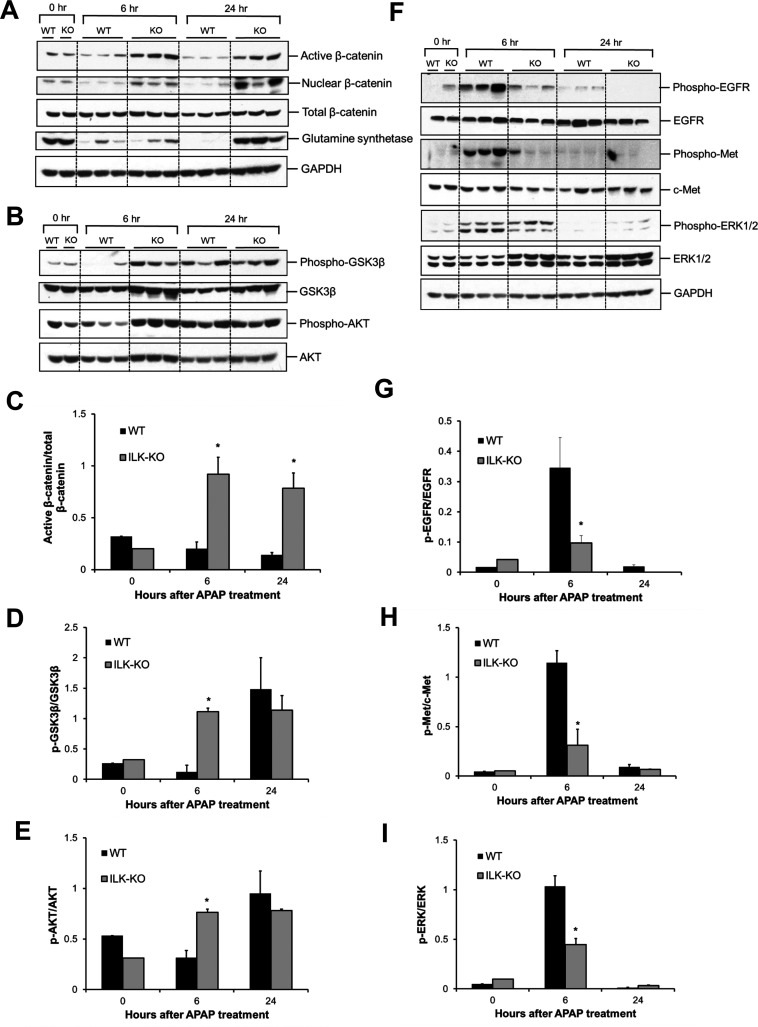
Figure 3.
Differential activation of proregenerative signaling after APAP overdose in ILK KO mice. (A) Western blot analysis of active β-catenin, nuclear β-catenin, total β-catenin, glutamine synthetase, (B) phospho-GSK3β, GSK3β, phospho-AKT, and AKT using total cell extract (unless specified) of liver of WT or ILK KO mice treated with 300 mg/kg APAP. Densitometric analysis showing (C) activation of β-catenin, (D) inactivation of GSK3β, and (E) activation of AKT, respectively, based on Western blot images shown in (A) and (B). (F) Western blot analysis of phospho-EGFR, EGFR, phospho-Met, c-Met, phospho-ERK1/2, and ERK1/2 using total cell extract of liver of WT or ILK KO mice treated with 300 mg/kg APAP. (G–I) Densitometric analysis showing quantification of expression proteins shown in (F). All samples were collected at 0, 6, and 24 h after APAP treatment. Significant difference between groups at *p < 0.05.

