Abstract
Crouzon syndrome (CS) is an autosomal dominant genetic disorder characterized by craniofacial dysostosis. Premature fusion of skull base leads to midfacial hypoplasia, shallow orbit, mandibular prognathism, overcrowding of upper teeth, high-arched palate, and upper airway obstruction. It is important for anesthesiologists managing such patients to recognize and avoid potential airway complications. Here, we present a case of a 10-year-old child with CS posted for ptosis correction surgery. Use of peripheral nerve blocks to cut down opioid requirement, inhalational induction, and maintenance are key aspects in successful management of such cases.
Keywords: Airway problems, Crouzon syndrome, inhalational induction, peripheral nerve blocks
INTRODUCTION
Crouzon syndrome (CS), named after the French neurosurgeon Octave Crouzon is an autosomal dominant genetic disorder characterized by craniofacial dysostosis.[1,2] Premature fusion of skull base leads to midfacial hypoplasia, shallow orbit, mandibular prognathism, overcrowding of upper teeth, high-arched palate, and occasional upper airway obstruction.[3] These patients can present for reconstructive surgeries and other unrelated surgeries. They pose a challenge for an anesthesiologist mainly due to difficult airway.[4,5] Here, we present a case of 10-year-old child with CS posted for ptosis correction surgery.
CASE REPORT
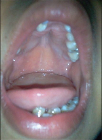
A 10-year-old male child, weighing 24 kg was posted for the left congenital ptosis correction (levator palpebrae superioris resection). He had previously been diagnosed to have CS by the pediatricians at our institute with features of facial dysmorphism such as dental crowding, enlarged adenoids, and deviated nasal septum to left side. Examination of the airway revealed a high-arched palate, with adequate mouth opening and neck movement [Figure 1]. The child had a history of delayed milestones and was of short stature. The parents gave a history suggestive of obstructive sleep apnea. The child had undergone adenotonsillectomy 1 year back at a peripheral center. Postsurgery the patient was kept in the pediatric Intensive Care Unit (ICU) for 1 day. Noninvasive ventilation was given to maintain the airway. There were no hospital records available and the parents failed to give any further detail.
Figure 1.

High-arched palate in the patient.
The parents were counseled before the surgery and consent taken for postoperative ICU stay. Anticipating a difficult airway, difficult airway cart comprising airways of all sizes, tracheostomy kit, laryngeal mask airways (LMAs), and fiberoptic bronchoscope were kept ready in the operating room (OR). No premedication was advised.
The child was kept first in the surgery list. Our plan of anesthesia was general anesthesia with minimal use of opioids and propofol. On shifting the child to the OR, routine monitors including electrocardiography, noninvasive blood pressure, peripheral capillary oxygen saturation, and end-tidal CO2 were connected. Twenty-two-gauge intravenous (i.v.) access was secured in the left hand. The child was preoxygenated with 100% oxygen for 3 min. Anesthesia was induced with titrated dose of sevoflurane and 0.5 μg/kg fentanyl. On confirmation of loss of verbal reflex, check-ventilation was done. Due to facial dysmorphism, chin lift and jaw thrust had to be applied to facilitate adequate mask ventilation. After deepening the plane of anesthesia with propofol 10 mg, the airway was secured with flexometallic LMA size 2.0. We avoided the use of muscle relaxant or any further top up of fentanyl. The child was maintained on intermittent positive pressure ventilation mode on combination of 50% oxygen, 50% air, and sevoflurane. Supraorbital and supratrochlear nerve blocks were given with 1 ml of 0.5% bupivacaine each.
Intraoperative period was uneventful. Supplemental analgesia in the form of i.v. paracetamol 15 mg/kg was administered. The child was extubated when fully awake and shifted to recovery room in lateral position. Postoperative monitoring was done for 2 h and thereafter, the patient was shifted to the ward.
DISCUSSION
CS is an autosomal dominant syndrome characterized by a triad of skull deformities, facial anomalies, and exophthalmos. CS has a prevalence of 1:60,000 live births with male to female preponderance of 3:1.[6,7] CS is caused by mutation in the fibroblast growth factor receptor 2 genes. This syndrome is characterized by premature synostosis of coronal and sagittal sutures leading to facial dysmorphism. These features either become more prominent or may regress over time. Hearing loss is common (55%) and there is 30% incidence of C2, 3 spinal fusion. Mental capacity is usually normal in these patients.[8]
The most challenging aspect in a case of CS is airway management during any surgery performed under general anesthesia. The goal of anesthesia is proper preoperative preparation, avoiding sedative drugs, use of peripheral blocks, and awake extubation. In a case described by Harde et al., attempts at securing the airway by LMA and endotracheal under general anesthesia by conventional techniques had failed. The patient had to be awakened from anesthesia and airway was secured using fiberoptic bronchoscope after airway topicalization.[9] Difficult intubation has also been described by Kim and Kim wherein they intubated the trachea using airway exchange catheter inserted through LMA Fastrach.[10] Our patient had history and features suggestive of difficult airway (high-arched palate, retrognathia, postoperative noninvasive ventilation). Difficult airway cart was kept ready. We avoided the use of preoperative sedation and muscle relaxant. Minimal use of fentanyl and propofol was done. Supratrochlear and supraorbital nerve blocks were given which provided adequate analgesia perioperatively.
Neuraxial anesthesia might be the anesthesia of choice in many surgeries. The presence of scoliosis and vertebral anomalies might make it technically difficult.[11] Use of peripheral nerve blocks is very useful in these patients. We blocked the supratrochlear and supraorbital nerve providing adequate analgesia and cutting down the need of opioids.
Hence, proper preoperative examination, inhalational induction, avoiding sedative premedication, and minimal use of opioids and awake extubation are key aspects in successful management of such cases. The role of peripheral nerve blocks to minimize opioid usage is paramount.
Financial support and sponsorship
Nil.
Conflicts of interest
There are no conflicts of interest.
REFERENCES
- 1.Gorlin RJ, Cohen MM, Levin LS. Syndromes of the Head and Neck. 3rd ed. Oxford: Oxford University Press; 1990. pp. 516–26. [Google Scholar]
- 2.Nargozian C. The airway in patients with craniofacial abnormalities. Paediatr Anaesth. 2004;14:53–9. doi: 10.1046/j.1460-9592.2003.01200.x. [DOI] [PubMed] [Google Scholar]
- 3.Hlongwa P. Early orthodontic management of Crouzon syndrome: A case report. J Maxillofac Oral Surg. 2009;8:74–6. doi: 10.1007/s12663-009-0018-7. [DOI] [PMC free article] [PubMed] [Google Scholar]
- 4.Hughes C, Thomas K, Johnson D, Das S. Anesthesia for surgery related to craniosynostosis: A review. Part 2. Paediatr Anaesth. 2013;23:22–7. doi: 10.1111/j.1460-9592.2012.03922.x. [DOI] [PubMed] [Google Scholar]
- 5.Järund M, Lauritzen C. Craniofacial dysostosis: Airway obstruction and craniofacial surgery. Scand J Plast Reconstr Surg Hand Surg. 1996;30:275–9. doi: 10.3109/02844319609056405. [DOI] [PubMed] [Google Scholar]
- 6.Bowling EL, Burstein FD. Crouzon syndrome. Optometry. 2006;77:217–22. doi: 10.1016/j.optm.2006.03.005. [DOI] [PubMed] [Google Scholar]
- 7.Horbelt CV. Physical and oral characteristics of Crouzon syndrome, Apert syndrome, and Pierre Robin sequence. Gen Dent. 2008;56:132–4. [PubMed] [Google Scholar]
- 8.Friedhoff RJ. Anesthesia for pediatric craniofacial surgery. Finnanest. 2000;33:387. [Google Scholar]
- 9.Harde M, Suryavanshi V, Chhatrapati S, Vaidyanathan M, Bhadade R. Crouzon syndrome: An anesthetic challenge. Ain Shams J Anesthesiol. 2008;8:683–5. [Google Scholar]
- 10.Kim YH, Kim JH. Tracheal intubation in a patent with Crouzon's syndrome using LMA-Fastrach [TM] with the cook airway exchange catheter. Anaesth Intensive Care. 2009;37:145–6. [PubMed] [Google Scholar]
- 11.Bajwa SJ, Gupta SK, Kaur J, Singh A, Parmar SS. Anaesthetic management of a patient with Crouzon syndrome. South Afr J Anaesth Analg. 2012;18:270–2. \. [Google Scholar]


