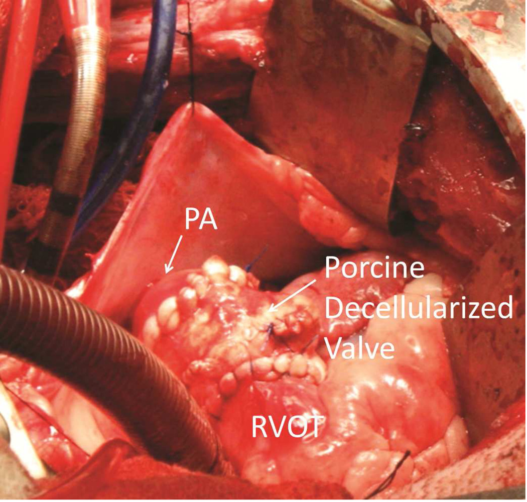
Figure 1.
Completed Pulmonary Valve Replacement in a Juvenile Sheep Model. The pericardium is shown reflected with silk sutures. Also visible are the venous and aortic cannulas for cardiopulmonary bypass. The porcine decellularized valve is shown after completion of both anastomoses. PA indicates pulmonary artery; RVOT, right ventricular outflow tract.

